Pages

Wednesday, December 7, 2016

Blog Post Week 10 12/02/16

This week the group needed to pour and run gels for the following samples:
Monday 205 lab (10 samples:  5 biofilm & 5 water)
Tuesday 205 lab (Cori 12 samples)
Wednesday day 205 lab (10 samples: 5 biofilm & 5 water)
Wednesday night 205 lab (12 samples:  6 biofilm & 6 water)

We also had to make gel maps in order to determine the number of gels we needed which we needed to pour gels once again on Thursday before our meeting. We made a total of 8 gels because we were screening multiple samples. We started to make the gels the day before to make it a little easier for the team and use our time more efficiently to progress on our data and project.
Displaying 20161202_121432.jpg
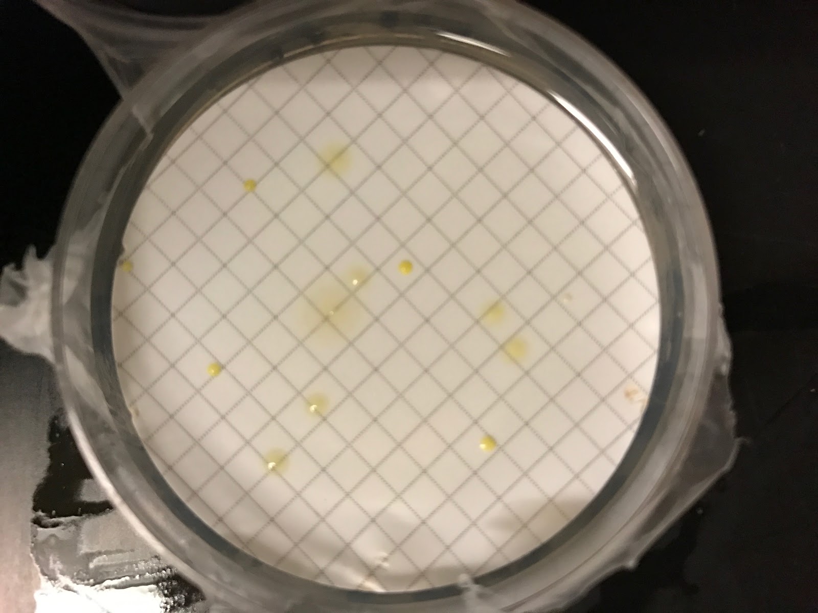
Sample of our gel using the Gel doc EZ imager that
shows us the bands of DNA and base pairs to see
which ones turned positive.

Displaying 20161130_154024.jpg
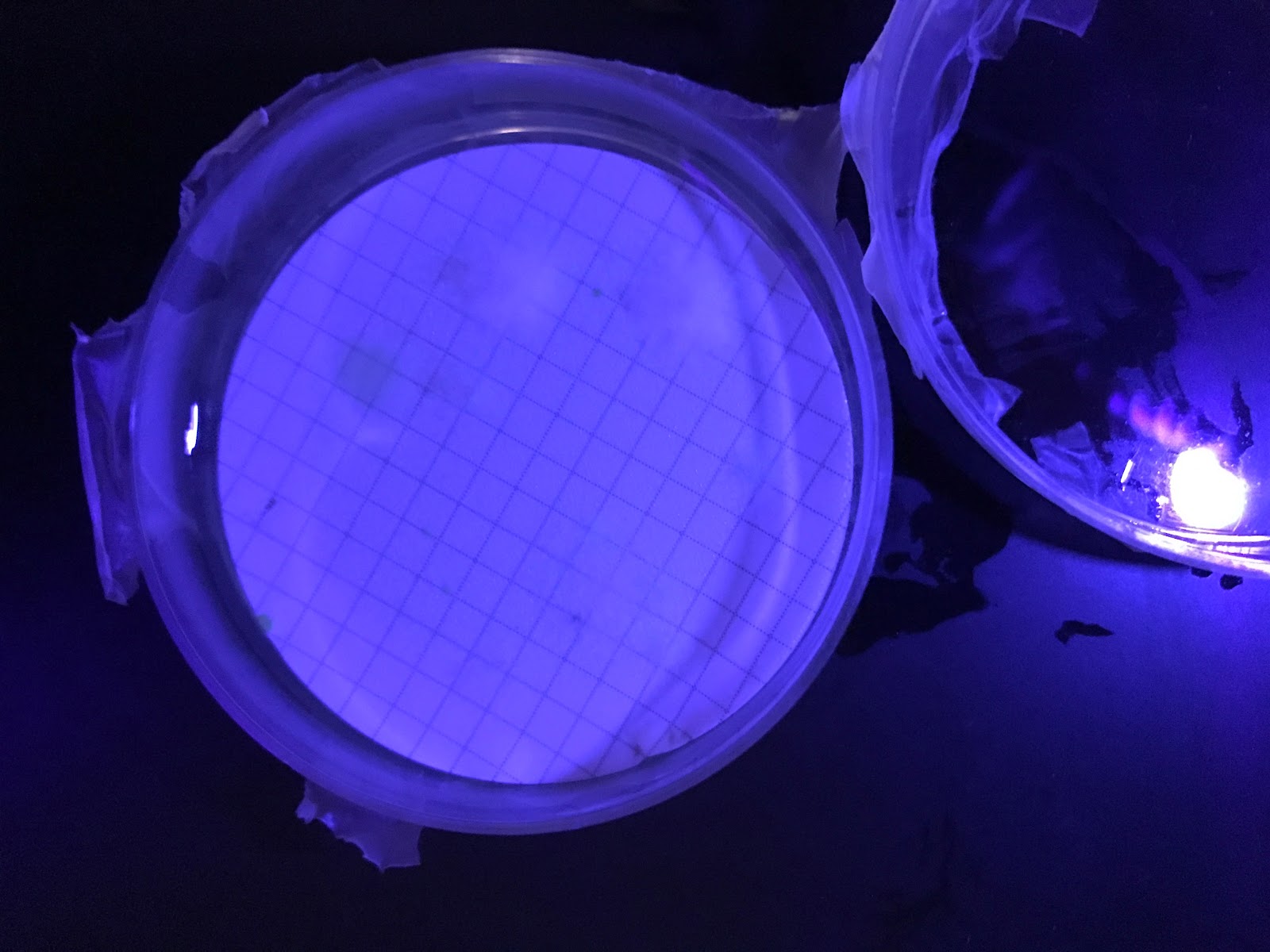
UV light through gels to see DNA

Blog Post Week 9 11/25/16

This week is Thanksgiving week so we did not meet on our regular scheduled day but did meet and come in during the weekday. The microbiology labs have done DNA extraction for their samples and we followed by nano dropping. The microbiology classes also ran a PCR and we were in charge of nano dropping that as well and screening them through gel electrophoresis.
This week the group set up PCR for the following samples:
Monday 205 lab (10 samples:  5 biofilm & 5 water)
Wednesday day 205 lab (10 samples: 5 biofilm & 5 water)

Wednesday night 205 lab (12 samples:  6 biofilm & 6 water)
Displaying 20161130_154110.jpg
Samples from the classes with their zones, sites and sources

Blog Post Week 8 11/18/16

This week we Nano dropped DNA samples from the Mon, Tues & Wed 205 labs before Friday. We also screened the gel results from the Mon, Tues & Wed 205 labs. The next step was determining which samples to run for DNA sequencing. Lastly, we began organizing data from Mon, Tues & Wed 205 labs into a table and created a composite of plate images from samples testing positive and the data should be organized by Zone, Site and Source. In addition, the microbiology classes have started collecting water and bio-films samples. Our team is now in charge of using the class samples and taking the data and compile it with ours. This week we took pictures of the plates and began to organize a file that will include the classes data. 
Displaying 20161130_154017.jpg
Some samples from the MicroLab courses.

Blog Post Week 7 11/11/16

Due to Veterans day, our research team did not meet, however, some of us throughout the week ran PCR once again and analyzed data to see if what we have has matched our previous results.

Displaying 20161130_154108.jpg

Blog Post Week 6 11/04/16

This week as a grade we were assigned for the independent research projects. We got to choose from three being: 1) Continuing collecting water samples, and completing steps as previously performed 2) Going to ASU West and using their equipment to find specific proteins 3) developing protocols for removing micro-plastics in the water using Pseudomonas. For the reason being that I am doing a project and poster for my honors class, and having a second person, I was required to stay with the 1st independent research project. It is also crazy to think that from here there is only 1 month left to collect and analyze water and bio-film samples for the project. We also analyzed the PIA and PF filtration plates from 4 lab sections and inoculated TSB broths for all samples testing positive.
D1HCWaterRefillWaterPIA.jpg
Pseudomonas Isolation Agar (PIA)
Pseudomonas Florescence (PF)

Blog Post Week 5 10/28/16

This week we poured and ran the gels for our positive controls, ran PCR on our positive controls and screened the positive controls to choose the best ones to create more positive controls to test the Pseudomonas (PA) and (PS) control samples. The PA samples are those of Pseudomonas aeruginosa and the PS samples of Pseudomonas species. We also learned how to do DNA extraction from the gels by cutting the bands out which is something new that I hadn’t done before nor did I perform this on my last project working with the protocols for E. coli. So definitely it was fun learning a new procedure that can be done to keep on confirming our DNA and positive controls.
Displaying 20161130_154144.jpg
Our tubes needed to perform PCR containing the primers.

Bog Post Week 4 10/21/16

This week for the Pseudomonas project we ran PCR on Pseudomonas aeruginosa DNA to create more positive controls for ourselves and the microbiology labs and as well performed PCR on the Pseudomonas control DNA using PA primers. So what is very interesting as well for this project and term is that three courses this semester are getting together to test the water and biofilm samples as well, which means more collection for us and more DNA data that we can use for consistency. This is a very first-time experience because the classes will have an opportunity do to real life undergraduate research and have that experience and a little taste to what researches do on a day to day basis. We also assisted with the “Creating Undergraduate Research Experiences Workshop,” which was for faculty only, but because we were part of the internship team, we got to help out, and therefore be a part of this great starting opportunity for undergraduate research at the community college level. 
Displaying 20161107_113311.jpg
The thermocycler  that allows for PCR to be ran by three important steps.

Blog Post Week 3 10/14/16

Continuing from last week the DNA extraction, was performed by following a protocol that was made during the summer by another intern. One way that we can quantitatively see and confirm how much DNA was actually in the tubes and shown in the gels is by Nano dropping. This shows a couple of numbers that let us know exactly what is DNA and what is just junk. Following the Nano drop, we proceeded to do PCR. PCR is an amplifier for our DNA using primers that are specific to the pseudomonas species we are looking for. However, there were troubles with the PCR because some of the samples didn’t have the full 25 microliters that were needed for the PCR so we had to rerun PCR on all of the samples. The goal was for the ones that turn out positive from the DNA gel extraction, then we are to send the positives to perform DNA sequencing at Arizona State University. 
Gels running for electrophoresis

Tuesday, December 6, 2016

Blog Post Week 2 10/07/16

This week for the Pseudomonas project we prepared and learned how to pour gels using the powdered agarose to create the gels to perform gel electrophoresis on our samples. (Since it is a team research project and Daisy and I are in this team and in STEM, feel free to look at both to learn more of what we do.) We have done water and bio-film collection from different sites and zones from our map. We have moved on to do filtration, using a filtration apparatus to trap any bacteria onto a filter. That filter was then transferred onto a plate to allow for us to culture the bacteria. The growth was then transferred into broth to allow to proceed into DNA extraction.
 
Screen Shot 2016-07-18 at 8.36.50 PM.png

Friday, September 30, 2016

Blog Post Week 1 (Pseudomonas Project)

Recap of what we have done with our Pseudomonas team: We had our introductions and have some new people on board for this semester. We started off by learning about Pseudomonas and its effects and then we learned how to do our primers and what gene we would need to look for. So far we have now collected water samples from various locations around campus and we filtered them, put them on a plate (PF, PiA) and grew the culture overnight. We then have inoculated from the growth of the plate into a broth and incubated that. Today we are in our process of conducting our first protocol and wait until next week to read the results and continue with our research.
Results of incubation from Mens
hot-tub
Filtered H2O from Mens hot-tub
and incubated


Wednesday, May 4, 2016

Blog Post Week 10

This week is a wrap up. I have been working on my final research paper and putting everything together of what I have done here in the internship for this semester. I have to say I have learned a lot from my mentors and peers. However, it is still not over. We still have one last presentation at Metro tech and I am pretty excited considering that I graduated from there. So hopefully I get to see some of my teachers as well. I just want to thank everyone who has been a part of this great experience being my first semester here in STEM. The knowledge and experience I have gained will help me in life and enhance my opportunities to continue doing research in the future. 
Nanodrop used to calculate the
amount of DNA and its purity.

Tuesday, May 3, 2016

Blog Post Week 9

This week was so crazy preparing for our presentations which were on Thursday. We went over our posters with our mentors and they gave us feedback on what we should say, how to say, what was on the rubrics, etc... It was a really great help to have them there always willing to help us and guide us through our project. During the conference we got to meet a lot of people and their research topics which was very interesting to hear. It was a really great experience and the food was great as well. Unfortunately I had to leave early because I had a Chemistry test and so I wasn't able to stay until the end. However, I heard that we did great as a school and our mentors are very proud of us. I know next year will be a successful one as this year and hopefully we have more students taking places for the conference.
These are my two gels before I ran then
 through Electrophoresis. I had two batches
and there were six protocols therefore I made
two gels with one batch on one and the second
batch on the other. The Yellowish/Orangish
color is the molecular ladder and the
blue are my samples with the
cyber-green on them. 

Sunday, April 17, 2016

Blog Post Week 8

Good afternoon everyone! Hope you are all having a great week. This wee was filled with lots of fun and work. I got to do my protocols once again and this time I was able to do them where I took half the time as last time. I learned how to choose carefully which steps to take so that I would save time. For example, if something needs to be centrifuged at 4000 rpm for 10 mins then I would see where another protocol I have to do the same and I'll do both together, instead of doing one at a time. This week I am also focusing on my template and will keep on editing as I go. With a few more weeks for the conference I will do one more set of data so that I can have 3 all together to present. Once again thank you to all who is making this possible and I look forward to keep on working here.


These are five of six protocols in which I am using to conduct my project.
(sixth one is a Kit, therefore it has its own protocol)

Monday, April 4, 2016

Blog Post Week 7

Good evening everyone! Still very excited to be able to be with you all at the conference. This week I have inoculated a new broth and my first samples were practice samples. I was still confused because my project is a bit different but that’s why we have mentors haha. Josh has been helping me and it’s been really helpful to have his insight. I have an idea now and all I need is the long periods of time so that I can do all my protocols at once in order to have a control. This would be having the same bacteria, from the same culture, right from when I take it out after being incubated. I have also re-learned how to use the spectrophotometer to calculate the concentration of my bacteria. 
This shows the spectrophotometer and my tubes that I will be using to
obtain my zero and then have my reading from my bacteria.
It will be set to 600 nm for a reading.

Blog Post Week 6

Good evening everyone! So this week I’m sure you guys found out if you were accepted and we all were! That is awesome! I can’t wait to be there with all of you and share these unforgettable experiences. Well this means that I have to start working on my project to have data for the presentation. I’m sure all of you know how Science goes but I’ve had to inoculate and re-inoculate my tubes because I need a fresh 24hr or less bacteria growth. However my time sometimes doesn’t allow me to come in to be under the 24hrs. However I have had some data and I have started using the pipettes and the micro centrifuge tubes. So I’m getting hands on and I’m learning so much more. Here are some of a few samples that I have been working on with the protocols.
As you can see I have lined my samples (1st row left-right) up
 with their finished products (1st column up-down) 

Blog Post Week 5

Good evening everyone! I’m pretty sure we were all excited this week to turn in our abstracts and see our results. Well this week I am now focusing on getting my protocols and making sure they are ready. Since my project will be a little different than others I need to get with my mentors and make sure that I am doing the correct procedures to make my experiments go right. I have also met with Matt and will discuss with him on some protocols from recent students that I can use and some that I will have to make up. These are some solutions that I will need in doing some of my protocols.
Lysis Buffer, NaOH Solution, Meat Tenderizer
Not in the picture:(TE Buffer, HCl Solution)

Thursday, March 10, 2016

Blog Post Week 4

Good afternoon everyone! So this week we were all working on our abstract and was the focus researching and getting background information that we need for this semester. Thank you to all the mentors and peers for their support as well. Honestly, it is hard to think about how you should word your abstract and trying to find what is the best, most vital information that should be included in the abstract.  However, we got it done and I’m anxious to see how well we do. On the other hand, I have also been working with David’s team and today I learned how to do water filtration so thanks to the team for showing me. I got to work under the hood and get a feel for what I have to do and will be doing in the future.

This is the hood that one must work in when doing water filtration.

Wednesday, March 2, 2016

Blog Post week 3


1. Oxidase Test[no color change(-)]
4. Found Unknown (E-Coli!)

Hey everyone! Good news! As you know I had made a mistake last week and had to regrow my cultures. Well they grew! This time I was able to determine under the microscope that my bacteria was a gram negative-bacilli. Using a flow chart, it guided me to the different tests I were to perform to find my unknown. The following three pictures show the results of my tests and the last one is my own flow chart that I made as I performed each test. The first test after identifying it was a gram-negative bacilli, was called the oxidase test. By doing this we determined that it was negative because by placing a drop on a colony, it would either have changed color if it was positive. Next were the glucose fermentation test and the SIM test that I had to leave incubated over a 24hr period. These are the results I found today. For the GFT, I found that it was acid because of the yellow color, and it was a gas by the bubble in the Durham tube. Since we had a positive acid, I was then directed to look at the SIM testing results which was a stab of my bacteria into the tube and found that there was a negative in the reduction of sulfide which if it was positive it would have turned a dark color. I also found that the motility was positive because as you can see, it looks as if it came away from the straight stab that I had made, therefore there was movement, in which it made the motility positive. By performing these tests I have found my unknown!Which is circled on the 4th picture.
2. GFT Yellow color and bubble(+ Acid)
3. SIM (-)Reduction of Sulfur and (+) Motility


Thursday, February 25, 2016

Blog post Week 2

Good afternoon everyone. So this is week 2 of S-STEM and I have learned very much about the procedures and how to classify bacteria. This week I took my TSA plates and saw that bacteria had multiplied. By this I was able to choose one single colony to begin my procedures in finding what kind of bacteria it is. Along the way, I made a simple error and unfortunately I have to redo it again. It was after putting them in the micro glass that adding the chemicals to help identify the characteristics of bacteria is where I made an error as. I noticed when I saw through them on the microscope that I couldn't identify them as either gram negative or gram positive because they were both colors. As a result, yesterday I began a new TSA plate and let it incubate for 24hrs. However, coming back today, I took it out and saw that nothing had grown on it! So I have redone now two TSA plates: One using the broth and the other using the actual dish that the bacteria was on. Hopefully by tomorrow I have the multiplied growth of bacteria to find my unknown.
Multiplied growth of unknown Bacteria
Micro glasses with my bacteria to view under a microscope


Wednesday, February 17, 2016

Blog Post Week 1

        Good afternoon everyone. It’s exciting to be part of S-STEM this semester and hope to complete a great internship. Well today I began my project by taking an unknown sample, and making what is called a lawn streak (Top) and an isolated streak (Bottom). The lawn streak is done to make a culture of the unknown bacteria, by the individual cells multiplying to form millions of identical cells. The three quadrant streak is to isolate single colonies of the unknown for macroscopic examination. They were placed in the incubator with a temperature of 36.7 degrees Celsius where the growth will begin. The ideal temperature for bacterial growth is 25-37 degrees Celsius.

Lawn Streak and Isolated Streak of Unknown (before)